Preparing for Dialysis Access Surgery
When hemodialysis may become necessary, creating a dependable way to access the bloodstream is an important part of treatment planning. Hemodialysis moves blood from the body to a dialysis machine, where waste and excess fluid are removed before the blood is returned. Because this process is repeated several times each week, the access site must provide steady blood flow and tolerate frequent needle placement.
An arteriovenous fistula or arteriovenous graft may be created for long-term dialysis access. Before choosing a location or type of access, the care team needs detailed information about the patient’s arteries and veins. A physical examination can reveal visible veins, pulse strength, swelling, and previous scars, but it may not show the full condition of vessels beneath the skin. Vessel mapping provides a closer look.
Nephrology Associates of Northern Illinois and Indiana (NANI), with practices in Berwyn and North Chicago, Illinois, and Bergen, New Jersey, helps patients understand the preparation involved in kidney replacement treatment. Knowing what vessel mapping evaluates and how the results influence surgical planning can make the process feel more understandable.
What Is Vessel Mapping?
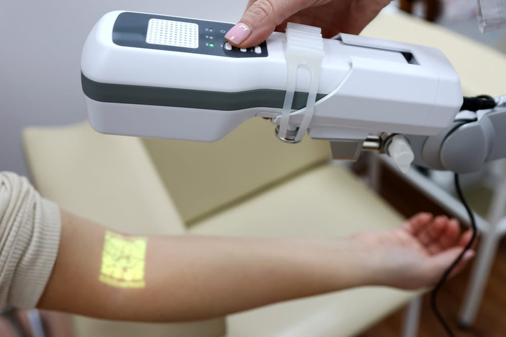
Vessel mapping is a noninvasive ultrasound examination used to evaluate the arteries and veins that may be considered for dialysis access surgery. The test is usually performed on the arms, although the exact area examined depends on the patient’s medical history and access needs.
During the examination, a vascular technologist applies ultrasound gel to the skin and moves a handheld transducer along the arm. The device uses sound waves to create images of the blood vessels. These images allow the care team to measure vessel diameter, evaluate blood flow, and identify possible narrowing, blockage, scarring, or clotting.
The technologist may examine both arms so the surgeon can compare potential access locations. A tourniquet or blood pressure cuff may be used briefly to help the veins expand and become easier to assess. The examination does not typically involve radiation, needles, incisions, or sedation.
The results become part of an individualized vascular access plan that considers vessel quality, treatment timing, previous procedures, and the patient’s broader health needs.
What the Ultrasound Measures
Vessel mapping evaluates both arteries and veins because successful dialysis access depends on adequate circulation through each. Arteries carry blood away from the heart, while veins return it. An AV fistula connects an artery directly to a vein, whereas an AV graft uses a synthetic tube to create that connection.
The ultrasound commonly measures the width of selected vessels. A vein must be able to enlarge and strengthen if it is going to mature into a usable fistula. The artery must also provide enough blood flow to support dialysis without creating circulation problems in the hand.
The test may also evaluate how deeply the veins lie beneath the skin. A vein that is healthy but positioned too deeply may be difficult to reach consistently with dialysis needles. Vessel pathway, branching patterns, and areas of previous damage can also affect surgical planning.
Blood flow is assessed to determine whether vessels appear open and whether circulation is moving in the expected direction. Prior IV lines, central venous catheters, blood clots, injuries, or surgeries may have affected veins that look normal from the outside.
These findings are interpreted together rather than used as isolated measurements. A single number does not determine whether a fistula or graft will succeed.
How Mapping Helps Plan an AV Fistula
An AV fistula is created by surgically connecting an artery to a nearby vein, most often in the arm. The increased arterial blood flow causes the vein to become larger and its walls to thicken. This process, called maturation, prepares the vein for repeated dialysis needle placement.
Because fistulas use the patient’s own blood vessels, they may offer long-term advantages when the anatomy is suitable. However, they require time to mature and do not always develop enough blood flow to support dialysis.
Vessel mapping helps the surgeon identify artery-and-vein combinations that appear most likely to mature successfully. It may reveal veins that are not easily visible or felt during a physical examination, expanding the range of options available for planning.
When possible, the care team may consider creating access farther down the arm before using vessels closer to the shoulder. This approach can help preserve additional sites for the future. The final decision depends on vessel size, circulation, medical history, treatment timeline, and the anticipated need for long-term access.
Mapping improves the information available before surgery, but it cannot guarantee that a fistula will mature or remain functional indefinitely. Ongoing monitoring is still necessary after placement.
How Mapping Supports AV Graft Planning
An AV graft connects an artery and vein using a synthetic tube placed beneath the skin. Dialysis needles are inserted into the graft rather than directly into a natural vein. A graft may be considered when the veins are not suitable for a fistula or when other medical factors make it the more practical option.
Even though a graft does not rely on a vein enlarging in the same way as a fistula, the surgeon still needs healthy vessels at both ends of the connection. Vessel mapping can help identify an artery capable of supplying adequate blood flow and a vein able to receive the returning blood.
The examination may also help determine where the graft could be positioned and whether previous scarring or vessel narrowing would interfere with placement. Grafts can often be used sooner than traditional fistulas, but they may have a greater tendency to develop narrowing, clotting, or infection.
The choice between a fistula and graft should reflect the patient’s overall treatment plan rather than one ultrasound result. Understanding how these options fit within kidney replacement therapy can help patients participate more confidently in access discussions.
Who May Benefit From Detailed Vessel Mapping?
Not every patient has the same vascular history or level of access risk. A physical examination remains important, but ultrasound mapping may provide additional value when suitable vessels are difficult to identify or when previous conditions may have affected circulation.
Factors that may make detailed mapping particularly useful include:
- Previous fistulas, grafts, dialysis catheters, or access surgeries
- A history of blood clots, arm trauma, or vascular procedures
- Veins that are difficult to see or feel during examination
- Diabetes, vascular disease, or circulation concerns
- Prior placement of IV lines or other devices that may have injured veins
- Swelling, differences in pulse strength, or concerns about blood flow
- Limited options because of vessel size or previous access failure
These factors do not automatically prevent fistula or graft creation. They indicate that closer evaluation may be helpful before selecting a surgical site.
What to Expect During the Appointment
Vessel mapping is generally a straightforward outpatient test. Patients may be asked to wear loose clothing or change into a gown so the technologist can access the arms. Watches, bracelets, and tight sleeves may need to be removed.
The patient usually lies on an examination table while the technologist evaluates one arm and then the other. Gel is placed on the skin, and the transducer is moved along the arm to trace the vessels. Mild pressure may be necessary to obtain clear images, but the test should not be painful.
The appointment length varies depending on the number of vessels examined and whether previous procedures have changed the patient’s anatomy. Once the examination is complete, normal activities can usually be resumed immediately.
The technologist records the images and measurements, while the surgeon or nephrology team reviews the findings in the context of the overall access plan. Results may therefore be discussed during a later appointment rather than at the time of the ultrasound.
Why Early Access Planning Matters
Access planning is often most successful when it begins before dialysis is urgently required. A fistula may need several weeks or months to mature, and some patients require additional procedures before the access can support treatment.
Waiting until dialysis must begin immediately can limit the available options. A temporary central venous catheter may be needed while a permanent access heals, and catheters carry different infection and maintenance considerations.
The timing of access planning is based on the rate of kidney function decline, symptoms, laboratory trends, medical history, and the likelihood that dialysis will be needed. Learning how kidney function changes over time helps explain why preparation may begin before kidney failure occurs.
Starting the conversation early gives patients time to understand treatment choices, complete vascular testing, meet with the surgical team, and prepare for recovery without the pressure of an urgent decision.
Protecting Blood Vessels Before and After Surgery
Patients who may need hemodialysis in the future are sometimes advised to protect the veins in their arms. Repeated blood draws, IV placement, and some vascular devices can damage or scar vessels that might otherwise be used for dialysis access.
Once an arm has been selected for access creation, the care team may recommend avoiding blood pressure measurements, routine blood draws, or IV placement in that arm. Instructions vary, so patients should follow their clinicians’ recommendations rather than assuming the same restrictions apply in every situation.
After a fistula or graft is created, daily monitoring becomes important. Patients may be taught to feel for the vibration known as a thrill, keep the site clean, and watch for swelling, redness, drainage, unusual pain, prolonged bleeding, or changes in blood flow.
These habits help identify potential complications early and support the long-term function of the access.
Moving Forward With a More Informed Access Plan
Vessel mapping gives the dialysis access team a detailed view of arteries and veins before surgery. By evaluating vessel size, depth, blood flow, and signs of previous damage, the test can help guide the selection of a fistula or graft location.
The ultrasound is one part of a larger decision. Physical examination findings, kidney disease progression, prior procedures, expected dialysis timing, and the patient’s long-term treatment goals all influence the final plan.
For patients preparing for hemodialysis, understanding why vessel mapping is performed can make the next steps easier to follow. Across its Berwyn and North Chicago, Illinois, and Bergen, New Jersey locations, NANI helps patients navigate dialysis preparation with attention to vascular health, treatment planning, and long-term access needs.
Sources
Lok, C. E., Huber, T. S., Lee, T., et al. (2020). KDOQI clinical practice guideline for vascular access: 2019 update. American Journal of Kidney Diseases.
Allon, M., Lockhart, M. E., Lilly, R. Z., et al. (2001). Effect of preoperative sonographic mapping on vascular access outcomes in hemodialysis patients. Kidney International.
Kosa, S. D., Al-Jaishi, A. A., Moist, L., & Lok, C. E. (2015). Preoperative vascular access evaluation for hemodialysis patients. Seminars in Dialysis.