Why Dialysis Access Monitoring Matters
A fistula or graft must provide reliable blood flow every time a patient receives hemodialysis. Even when an access has been working well, changes can develop over time. Narrowing inside a blood vessel, changes in pressure, clot formation, or problems with the surrounding tissue may affect how effectively blood moves through the access.
Monitoring is the routine process of checking how the access looks, feels, sounds, and performs during dialysis. These observations can provide important clues before an access stops working completely. Patients, dialysis staff, nephrologists, nurses, and vascular access specialists may all contribute to this process.
Nephrology Associates of Northern Illinois and Indiana (NANI) supports patients receiving kidney care in Berwyn and North Chicago, Illinois, and Bergen, New Jersey. Understanding what is normal for an individual fistula or graft can make changes easier to recognize and discuss with the care team.
What Healthy Blood Flow Feels Like
One of the simplest ways to monitor an arteriovenous fistula or graft is by checking the thrill. A thrill is the vibration created as blood moves through the access. Patients are often encouraged to gently place their fingers over the access regularly so they become familiar with its usual sensation.
The access also creates a sound called a bruit. Healthcare professionals can listen to this sound with a stethoscope during a physical examination. Changes in either the thrill or bruit may suggest that blood flow is no longer following its usual pattern.
A healthy access should have a consistent pattern that becomes familiar over time. A thrill that becomes weaker, disappears, feels unusually strong or pulsatile, or changes noticeably deserves attention.
Learning the basics of vascular access can help patients understand why these simple daily observations are an important part of protecting an access used several times each week.
Clinical Monitoring During Dialysis
Dialysis staff evaluate the access each time it is used. Before inserting needles, they may inspect the skin, feel the access, and note swelling, tenderness, redness, or changes in the vessels.
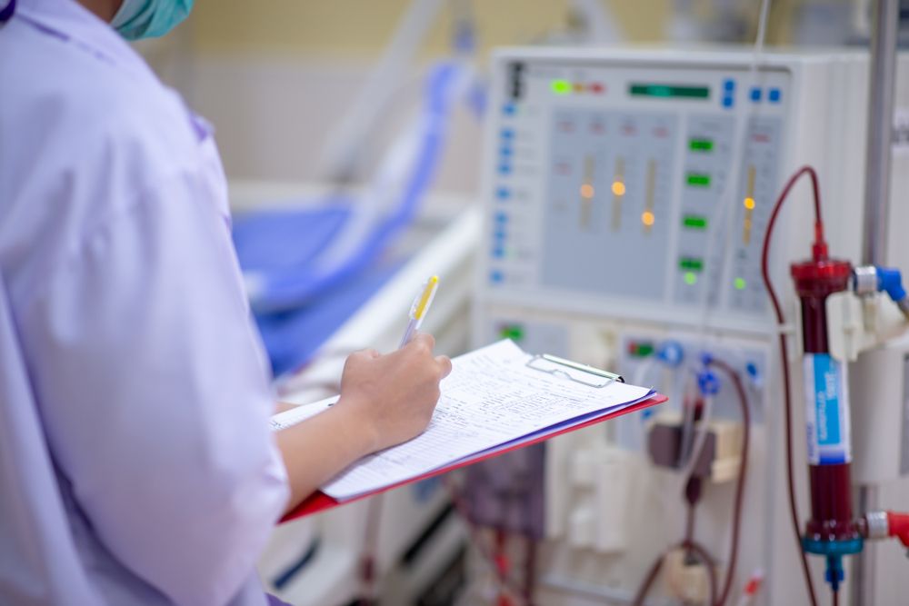
The dialysis machine also provides information. Blood-flow rates, pressures, alarms, and the effectiveness of the dialysis session may change if the access is developing a problem.
One unusual reading does not necessarily indicate access failure. Needle position, patient movement, equipment issues, or temporary changes can affect treatment. Patterns that occur repeatedly are more important because they may point to a developing blood-flow problem.
Clinical monitoring is therefore based on multiple observations rather than a single test or machine reading.
Signs That Blood Flow May Be Changing
Stenosis, or narrowing, can reduce blood movement through a fistula or graft. If the narrowing becomes significant, it may eventually contribute to clotting or interfere with dialysis.
Changes that may prompt further evaluation include:
- A weaker, absent, or noticeably different thrill
- Difficulty reaching the prescribed dialysis blood-flow rate
- Repeated pressure alarms during treatment
- Increasing difficulty inserting dialysis needles
- Swelling in the access arm or hand
- Prolonged bleeding after needles are removed
- Changes in the shape, firmness, or pulse of the access
- An unexplained decline in dialysis effectiveness
These findings do not identify the exact cause on their own. Their purpose is to signal that the access may need closer examination.
A sudden loss of the thrill or inability to use the access for dialysis requires prompt communication with the dialysis or vascular access team.
How Narrowing Can Affect an Access
Many access problems begin with stenosis. Narrowing can develop where vessels connect, within sections exposed to repeated needle placement, or in veins farther from the access.
As the opening becomes smaller, resistance to blood flow increases. Pressure may build within the access, and dialysis staff may notice changes during treatment. Blood may also move more slowly through certain areas, increasing the risk of clot formation.
A stenosis does not always cause obvious symptoms outside the dialysis unit. This is why routine examination and tracking treatment performance are valuable.
Physical examination remains a major component of access monitoring because changes in pulse, thrill, bruit, arm swelling, and bleeding can provide evidence of clinically meaningful stenosis.
Monitoring Versus Access Surveillance
The terms monitoring and surveillance are related but describe somewhat different approaches.
Clinical monitoring involves routine observation and physical examination. It includes listening to what the patient reports, inspecting the access, feeling the thrill and pulse, and reviewing dialysis performance.
Surveillance generally refers to specialized testing that measures access function, such as access blood-flow measurements or duplex ultrasound.
Current recommendations place strong emphasis on clinical monitoring. Specialized surveillance tests can provide additional information, but abnormal test results alone do not always mean a procedure is needed. Research has shown that surveillance can identify stenosis, but its ability to predict which accesses will actually clot is less certain.
The decision to obtain imaging or perform an intervention is therefore usually based on the complete clinical picture.
When Ultrasound May Be Used
Duplex ultrasound can help evaluate an access when the physical examination or dialysis performance raises concern. The test uses sound waves to examine blood vessels and measure blood flow without requiring an incision.
Ultrasound may identify narrowing, clot formation, abnormal vessel enlargement, or reduced blood flow. It can also help evaluate a fistula that is not maturing as expected after creation.
Not every dialysis access requires routine ultrasound on a fixed schedule. Testing is often most useful when there is a clinical reason to suspect a problem.
If imaging confirms significant stenosis that is affecting access function, the care team can then determine whether treatment is appropriate.
Protecting the Access Between Dialysis Sessions
Monitoring continues outside the dialysis clinic. Patients can become familiar with the appearance and sensation of their access and report changes rather than waiting until the next treatment becomes difficult.
The access arm should generally be protected from unnecessary pressure. Blood pressure measurements, routine blood draws, tight clothing, and heavy straps over the access may be discouraged.
Patients should also watch the skin for redness, warmth, drainage, open areas, or unusual swelling. Infection can interfere with access function and may become serious if it spreads.
These daily habits are especially important because hemodialysis depends on a dependable route for moving large volumes of blood between the body and the dialysis machine.
As one form of kidney replacement therapy, hemodialysis cannot be delivered effectively when the vascular access does not provide sufficient blood flow.
What Happens When Monitoring Finds a Concern
An abnormal finding does not automatically mean that an access must be replaced. Many problems can be evaluated and, when appropriate, treated while preserving the existing fistula or graft.
The healthcare team may perform a more detailed physical examination, review dialysis-machine data, or order ultrasound or angiography.
If significant narrowing is found, angioplasty may be used to widen the affected vessel. A clot may require thrombectomy or another procedure to restore blood flow. Infection, aneurysm formation, poor maturation, or hand-circulation problems require treatment based on the specific condition.
The goal is not to perform procedures simply because a narrowing exists. Treatment is generally considered when clinical signs indicate that the problem is affecting access function or creating another meaningful concern.
Why Dialysis Performance Provides Useful Information
The purpose of a vascular access is ultimately to support effective dialysis. Changes during treatment can therefore reveal problems that may not be obvious when looking at the arm alone.
Blood must move from the patient to the dialysis machine at an adequate rate and then return safely. If access flow declines, dialysis may become less efficient, treatments may take longer, or machine alarms may occur more frequently.
Laboratory testing and dialysis adequacy measurements can help determine whether treatment is removing enough waste and fluid.
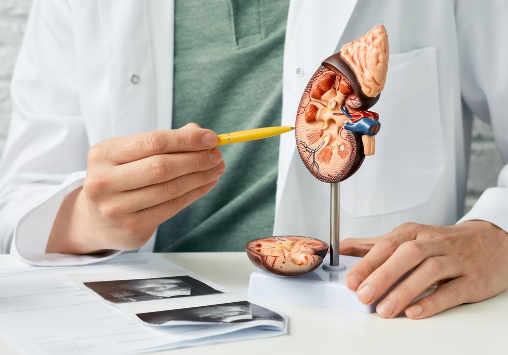
Understanding kidney function also provides context for why maintaining adequate dialysis blood flow is so important. When the kidneys can no longer perform sufficient filtration on their own, the dialysis treatment and the access that supports it become closely linked.
Keeping Blood Flow Reliable Over Time
Dialysis access monitoring works best when patients and healthcare teams understand what is normal and pay attention when that pattern changes. A difference in thrill, swelling, bleeding, needle placement, pressure readings, or dialysis performance may provide an early reason to investigate further.
No monitoring method can prevent every episode of stenosis or clotting. Routine physical examination and attention to treatment performance, however, can help identify clinically significant changes and guide decisions about additional testing.
For patients receiving kidney care through NANI in Berwyn and North Chicago, Illinois, and Bergen, New Jersey, maintaining a functional vascular access remains an important part of ongoing hemodialysis care. Consistent monitoring helps keep the focus on reliable blood flow and effective treatment rather than waiting until an access can no longer be used.
Sources
Salman, L., & Beathard, G. (2013). Interventional nephrology: Physical examination as a tool for surveillance for the hemodialysis arteriovenous access. Clinical Journal of the American Society of Nephrology.
Lok, C. E., Huber, T. S., Lee, T., et al. (2020). KDOQI clinical practice guideline for vascular access: 2019 update. American Journal of Kidney Diseases.
Valliant, A., & McComb, K. (2015). Vascular access monitoring and surveillance: An update. Advances in Chronic Kidney Disease.